
Visual Field Test (Perimetry) in Ahmedabad
A Visual Field Test (Perimetry) is an essential part of a Comprehensive Glaucoma Evaluation, measuring both your central and peripheral vision to detect early vision loss caused by glaucoma. Since glaucoma usually affects side vision first, regular visual field testing helps identify changes before symptoms become noticeable.
At Puja Eye Hospital, Naranpura, Ahmedabad, Dr. Puja Sheth, a trusted Glaucoma Specialist in Ahmedabad and Best Glaucoma Surgeon in Ahmedabad, uses advanced Visual Field Testing to diagnose glaucoma, monitor disease progression, and evaluate the effectiveness of your Glaucoma Treatment in Ahmedabad. Combined with modern diagnostic technology, this test helps protect the optic nerve, preserve vision, and support long-term glaucoma management.
What Is a Visual Field Test?
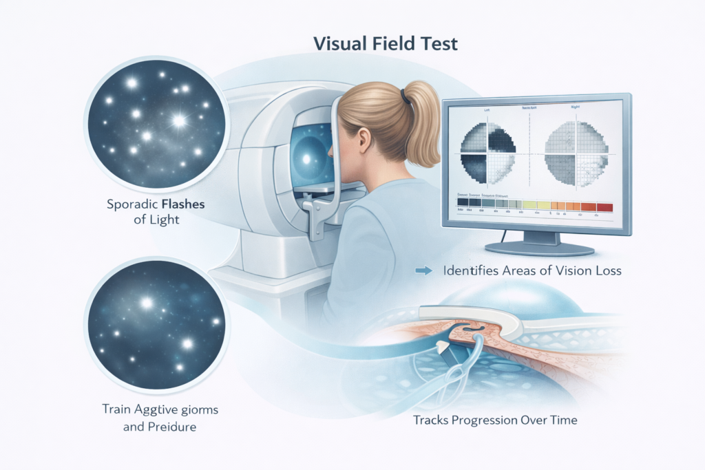
During a visual field test (perimetry), you sit in front of a specialised machine and look straight ahead at a central fixation point. At various locations across your field of vision, brief flashes of light appear at different intensities. Each time you see a flash, you press a button to indicate you have detected it.
The machine maps out your entire visual field and identifies any areas where sensitivity is reduced – indicating spots of vision loss. The result is a detailed map of your visual field, with statistical analysis comparing it to normal values for your age.
Why Is Perimetry Important in Glaucoma?
Glaucoma causes progressive damage to the optic nerve. As nerve fibres are lost, corresponding areas of the visual field become less sensitive or disappear entirely. The pattern and extent of visual field loss provides critical clinical information:
- It confirms whether functional (visual) damage is present, in addition to structural (nerve fibre) damage seen on OCT
- It establishes the current severity of glaucoma - mild, moderate, or advanced
- It detects whether glaucoma is progressing despite treatment, prompting a change in management
- It tracks the rate of progression - slow, moderate, or fast - which guides the aggressiveness of treatment
- It documents the patient's visual functional status for long-term clinical records
How Often Is the Visual Field Test Required?
The frequency of visual field testing depends on your individual situation:
- Newly diagnosed patients - typically 2 to 3 tests in the first year to establish a reliable baseline, as individual test results can vary
- Stable glaucoma - usually once or twice per year, depending on severity
- Progressing glaucoma or newly adjusted treatment - more frequent testing, sometimes every 3 to 4 months
- Glaucoma suspects (high-risk individuals without a confirmed diagnosis) - typically once a year
Consistent, regular perimetry over years is far more valuable than a single test. It is the pattern of results over time – the trend – that tells the most important story about whether glaucoma is being controlled.
Is the Visual Field Test Difficult?
The visual field test requires concentration and sustained attention, and some patients find it tiring, particularly in the first few attempts. It is important to understand that some variability between tests is entirely normal and expected – it does not mean your glaucoma has suddenly worsened.
With practice, patients become more comfortable with the test and results become more consistent. Dr. Sheth always reviews visual field results in the context of multiple tests over time, never making clinical decisions based on a single result in isolation.
The test takes approximately 5 to 10 minutes per eye.
Who Should Have a Visual Field Test?
- All patients diagnosed with any type of glaucoma, as part of routine monitoring
- Glaucoma suspects - patients with elevated eye pressure, suspicious optic nerves, or thin corneas - as part of baseline evaluation
- Patients with a family history of glaucoma who are being monitored annually
- Patients who have noticed any change in their peripheral vision or difficulty in low-light conditions
- Patients being evaluated for neurological conditions that can affect the visual pathway
Role of Visual Field Testing / Perimetry in Neurological Conditions
Visual field testing plays a vital role beyond glaucoma – it is an essential diagnostic tool in neurology and neuro-ophthalmology. The visual pathway travels from the eye through the optic nerve, optic chiasm, optic tracts, and ultimately to the occipital cortex at the back of the brain. Any disruption along this entire pathway – whether from a tumour, stroke, demyelination, or raised intracranial pressure – produces a characteristic pattern of visual field loss that helps localise the site of neurological damage.
- Key neurological conditions diagnosed or monitored with perimetry include:
- Pituitary tumours / Pituitary adenoma - Classic bitemporal hemianopia (loss of both outer visual fields) is the hallmark finding, caused by compression of the optic chiasm. Regular perimetry is critical for monitoring tumour growth and response to treatment.
- Stroke (Cerebrovascular Accident) - Occipital or parietal lobe strokes produce homonymous hemianopia (loss of the same side of vision in both eyes), which perimetry precisely maps and documents.
- Intracranial tumours & space-occupying lesions - Tumours pressing on the optic tract or radiations produce predictable field defects that indicate tumour location.
- Idiopathic Intracranial Hypertension (IIH / Pseudotumour Cerebri) - Raised intracranial pressure causes papilloedema and progressive peripheral field loss; perimetry is key to monitoring whether treatment is protecting vision.
- Multiple Sclerosis (MS) & Optic Neuritis - Demyelination of the optic nerve causes central scotomas or altitudinal defects; field testing tracks recovery and disease progression.
- Occipital lobe epilepsy & migraine with aura - Transient or permanent visual field defects can be mapped and documented.
Because each neurological condition produces a distinct and recognisable pattern of field loss, perimetry serves as a neurological “fingerprint” – guiding diagnosis, confirming the level of lesion, and tracking response to neurosurgical or medical intervention. Dr. Puja Sheth works in close collaboration with neurologists and neurosurgeons to provide precise, reliable perimetry reports that support multidisciplinary patient care.
If you have been referred for a visual field test by a neurologist, neurosurgeon, or physician – or if you have noticed asymmetric vision changes, tunnel vision, or visual disturbances – contact Puja Eye Hospital for a prompt evaluation.
Dr. Puja Sheth -Glaucoma Specialist
Dr puja Eye Clinic - Dr Puja Sheth
- +91 8780012121
- 401-402, The 132 Offices and Showrooms Near Shell Petrol Pump, Besides Indraprastha Saptak 132 Ft. Ring Road, AEC Cross Rd, Naranpura, Ahmedabad, Gujarat 380013
-
Monday to Saturday 9:30 am – 4:30 pm
Prior appointment necessary
Chief Glaucoma Consultant and Surgeon, Dr. Nagpal's Retina Foundation, Ahmedabad
- 079-22865537
- Retina Foundation Near the Underbridge, Old Raj Bhavan Road, Shahibaug, Ahmedabad - 380 004, Gujarat, India.
-
Opening Hours Mon to Friday 09:am to 05:pm
Sat – 09:am to 11:am
FAQs
No. The visual field test is completely non-invasive and painless. You simply sit in front of the machine and respond to light stimuli. No eye drops, instruments, or contact with the eye are involved.
Missing some flashes is completely normal and expected – no one detects every single stimulus. The test uses statistical analysis to distinguish between normal variability and genuine areas of reduced sensitivity. Dr. Sheth reviews the full statistical report, not just the individual responses.
Yes. You should wear your glasses or contact lenses for the visual field test if you normally use them for near or distance vision. Please bring your glasses to the appointment.
Yes. A single normal visual field test is reassuring but is not a guarantee that glaucoma is absent or stable. Glaucoma is a progressive condition that requires monitoring over time. Regular repeat testing is what allows early detection of any progression.
No. The visual field test is one component of a comprehensive evaluation. Glaucoma is diagnosed based on a combination of optic nerve appearance, OCT findings, eye pressure, and visual field results. No single test is sufficient for diagnosis.
A visual field test, also called perimetry, is a diagnostic test that maps the full extent of your field of vision – both central and peripheral- by measuring your ability to detect light stimuli at different locations in your visual field. In glaucoma, damage to the optic nerve produces characteristic patterns of visual field loss (arcuate scotomas, nasal steps, altitudinal defects) that are precisely documented by perimetry. Perimetry is the primary functional test in glaucoma management: while OCT measures structural nerve damage, visual field testing measures whether that structural damage has translated into functional vision loss. Both are essential for complete glaucoma assessment at Puja Eye Hospital, Naranpura, Ahmedabad.
Puja Eye Hospital uses the Humphrey Field Analyser – the global standard for glaucoma perimetry – for all visual field testing. The Humphrey Field Analyser uses the SITA (Swedish Interactive Thresholding Algorithm) testing strategy, which provides reliable, reproducible results in a shorter test time compared to older full-threshold strategies. Dr Puja Sheth uses 24-2 and 30-2 test patterns for standard glaucoma monitoring, and 10-2 central field testing for advanced glaucoma where central vision is at risk. Reliable Humphrey visual field results are the cornerstone of glaucoma staging and treatment decision-making.
The visual field test is performed in a quiet, darkened room. You sit in front of a bowl-shaped white screen (the perimeter), rest your chin on a support, and fix your gaze on a central target. A series of light dots of varying brightness are flashed at different positions in the visual field. Each time you see a flash, you press a button. The test assesses each eye separately and takes approximately 5–8 minutes per eye using the SITA-Fast strategy, or 8–12 minutes with SITA-Standard. Many patients find the first test challenging – it is a concentration-intensive test – but performance typically improves with practice. Dr. Puja Sheth’s team provides clear instructions and ensures patients feel comfortable before the test begins.
Standard automated perimetry detects functional vision loss only after approximately 25–35% of retinal ganglion cell axons have been lost – meaning early glaucoma may be invisible on perimetry. This is why Dr. Puja Sheth combines visual field testing with structural imaging (Zeiss OCT of the RNFL and optic nerve head) in every evaluation. OCT can detect structural nerve damage before field defects appear, enabling earlier intervention. Once glaucoma is established, however, visual field progression is the most important indicator that treatment is insufficient and needs to be escalated. Serial visual fields over time – analysed with trend and event-based progression analysis – are the backbone of long-term glaucoma management.
The frequency of visual field testing depends on glaucoma severity and stability. Standard glaucoma care guidelines recommend at least two visual field tests per year for most patients with established glaucoma. For patients with rapidly progressing glaucoma or those in whom treatment has recently changed, testing every 3–4 months may be appropriate to reliably detect change. For stable, well-controlled early glaucoma, one to two tests per year is adequate. At Puja Eye Hospital, Ahmedabad, Dr. Puja Sheth tailors the testing schedule to each patient’s risk profile, ensuring that progression is detected promptly and treatment escalated before significant functional loss occurs.
Humphrey visual field results are presented as a numerical map showing sensitivity at each tested location, a grey-scale map showing the pattern of loss visually, and a probability map indicating which areas are abnormal compared to age-matched normals. Key summary indices include Mean Deviation (MD), which indicates overall field loss severity (values more negative than -6 dB indicate moderate loss; more negative than -12 dB indicate severe loss), and Pattern Standard Deviation (PSD), which reflects the pattern of localised loss. Reliability indices – fixation losses, false-positive and false-negative rates – indicate test quality. Dr. Puja Sheth interprets results in the context of the full clinical picture, including OCT, IOP history, and prior field tests, not in isolation.
The visual field test is completely safe, non-invasive, and involves no radiation or injections of any kind. It uses only flashes of light and your responses. No drops are required for the test itself, although pupils may be dilated if the test is being performed as part of a broader evaluation on the same visit. The test carries no risks or side effects. It can be safely performed in pregnant women, children (with modified protocols), and patients with systemic conditions. The only requirement is that you can sit still, hold your gaze steady, and respond to light stimuli – the test coordinator will guide you through each step.
Visual field testing using the Humphrey Field Analyser is available at Puja Eye Hospital, Naranpura, Ahmedabad, under the supervision of Dr. Puja Sheth. Tests are performed by trained ophthalmic technicians following a standardised protocol to ensure data quality. Results are reviewed and interpreted by Dr. Puja Sheth in the context of your full clinical picture. The clinic accepts both self-referred patients and those referred by other eye specialists or physicians across Ahmedabad and Gujarat. Appointments can be scheduled in advance; urgent cases presenting with symptoms of acute angle closure or sudden vision change are prioritised.